ELECTRONIC MICROSCOPY PRODUCTS
TESCAN UniTOM XL
SPECTRAL
A New Dimension for Micro-CT
SPECTRAL CT provides contrast where conventional micro-CT cannot, and reveals information about the composition of a sample by looking at the entire x-ray spectrum used to image a sample.
SPECTRAL CT reveals this chemical information at any point within a sample in a non-destructive manner and combines it with high-quality structural information in the same sample.
For the first time in a commercial micro-CT system, we can acquire chemical data such as identifying precious metals for mining and recycling, differentiating between different polymers in engineered samples, or optimizing contrast in biological samples.


Discover More With CT SPECTRAL

K-Edge Detection
Absolute identification of elements without prior knowledge.Using k-edge detection, precious metals and elements can be identified without any knowledge of the sample.

TrueContrast™
Provides contrast where conventional CT cannot. When conventional CT cannot differentiate between materials, SPECTRAL CT performs multienergy analysis in a single scan.

Spectrum Matching
A treasure trove of information at your fingertips.Compare spectra to a reference database or to each other to gain information about composition, concentration, and density.
Applications

Spectral CT in the world of electronics

Classify polymers based on their chemistry

Non-destructive 3D elemental analysis
SPECTRAL Information Beyond the Incredible Image Quality of TESCAN UniTOM XL
TESCAN SPECTRAL CT is a complement to TESCAN UniTOM XL, TESCAN‘s high-volume, versatile, multi-scale micro-CT system. Adding SPECTRAL CT imposes no compromise on conventional CT imaging, and all standard TESCAN UniTOM XL functions remain available.
SPECTRAL Suite
Powerful Analytical Tools for SPECTRAL Data
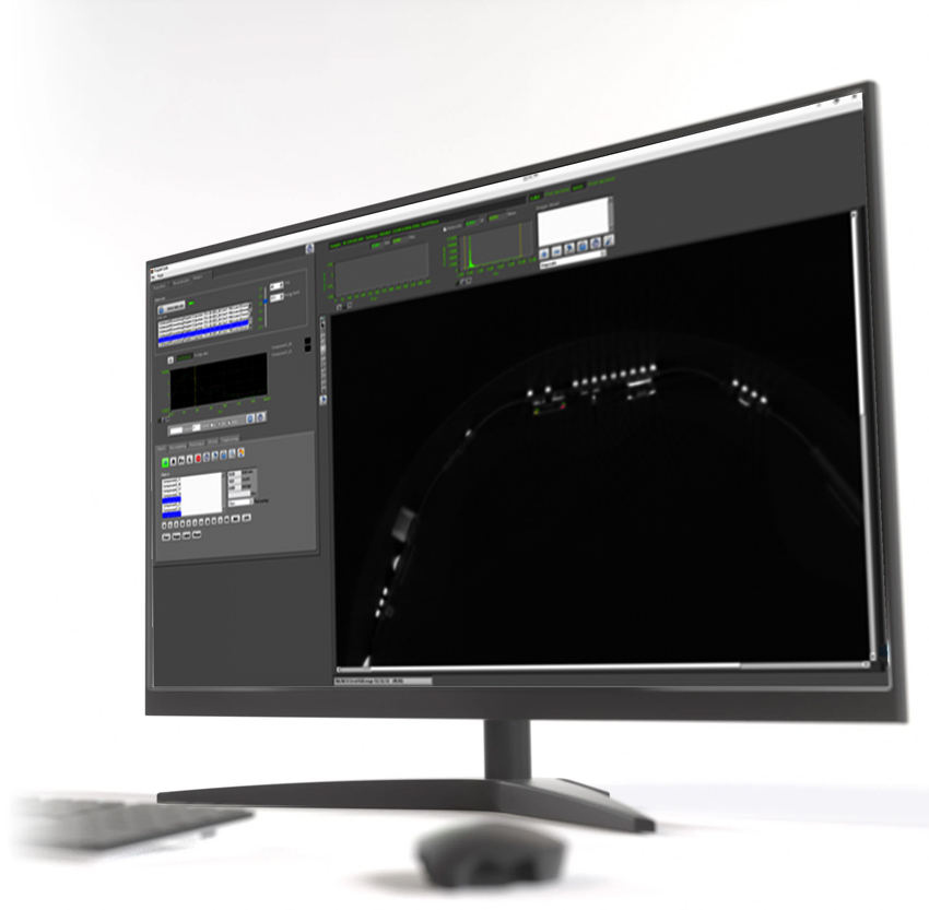
The TESCAN SPECTRAL suite exposes the power of PolyDET II, providing tools for SPECTRAL data acquisition, reconstruction and analysis. Through direct communication with AcquilaTM, TESCAN’s micro-CT control software, data can be acquired at any point within the sample using an intuitive VOI scanning workflow.
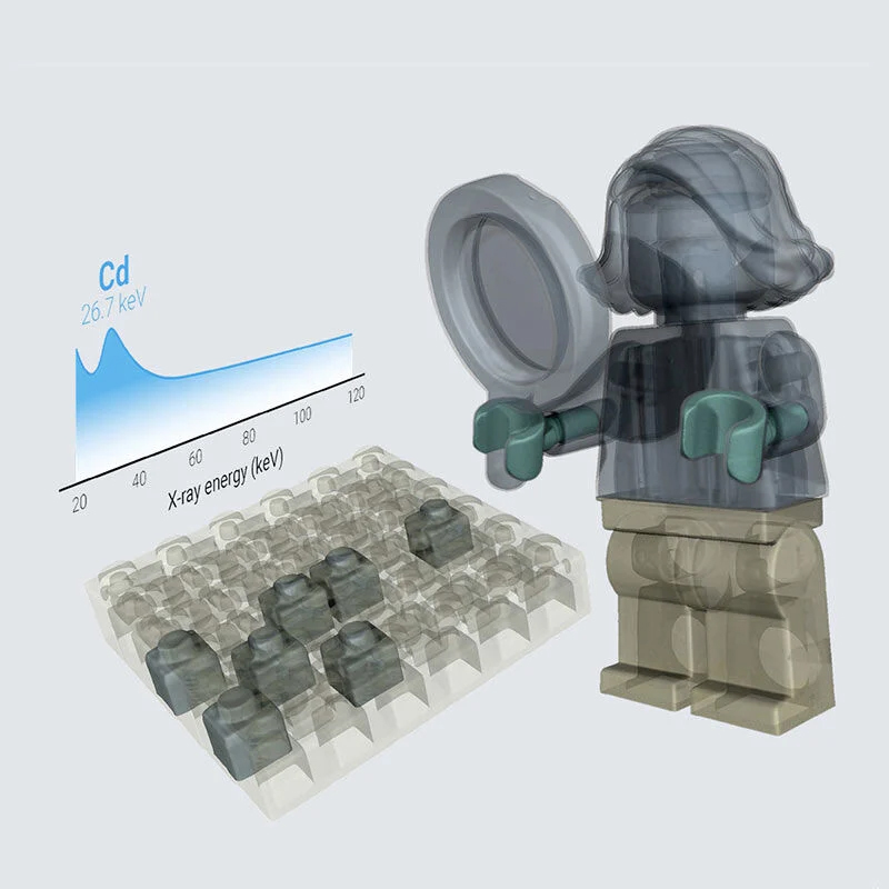
The reconstructed spectra can be visualized, analyzed and compared, enabling advanced analytical tools such as multi-energy CT, k-edge detection or atomic number plot extraction.

TESCAN UniTOM XL SPECTRAL At the University of Pau
The first installation of a TESCAN UniTOM XL SPECTRAL was carried out at the University of Pau (France). The Prof. Dr. Peter Moonen uses the system for multimodal characterization of rocks and metals and is impressed with the capabilities of TESCAN UniTOM XL.
The combination of conventional imaging and micro-CT enables a wide range of new applications for energy and mining applications.
Need more information?
Download Tescan SPECTRAL brochure
If you want more information about this equipment you must fill out the following form. Once completed, a direct download link will open.