
ELECTRONIC MICROSCOPY PRODUCTS
3D reconstruction and tomography
There are three ways to perform a 3D tomography on samples that are normally observed with an electron microscope, two are using the SEM itself directly and the third is by sequentially combining it with a high-resolution tomograph or micro-CT.
SBF reconstruction of a 3D volume in biological materials with the aid of an Ultramicrotome performing a reconstruction of serial sections or Serial Block Facing (SBF). The ultramicrotome can be inside or outside the SEM, thus speaking of in situ and ex-situ SBF. The technique of combining serial ultramicrotomy sections with the resolution and capabilities of a field emission microscope is a fast and affordable way to perform 3D reconstructions on biological samples with nanometer resolution.
Tissue and cell samples are embedded in resins and stained with heavy metals to be processed in an ultramicrotome and obtained images at low acceleration voltages.
The microtome can be incorporated into the TESCAN FESEM taking advantage of its special giant chamber specially designed to house the ultramicrotome or it can be left outside the microscope and follow a more laborious procedure where we preserve serial sections after passing through the FESEM. Images of each slice are aligned and stacked into a 3D package and later rendered using powerful 3D reconstruction software.
This software process is similar to what we would follow if we used a FIB to remove material layer by layer and obtain a high-resolution tomography.
FIBs have an ion column with which the sample can be nanosculpted and an SEM electron column with which an image of the resulting surface can be made. Thus, 3D reconstruction of a sample volume can be achieved by serial sections using FIB in combination with high-resolution SEM images of each cross section.
Once processing is complete, the 3D volume can be freely manipulated for visualization, and with additional treatments, such as thresholding or segmentation, it is possible to keep only some parts of interest and remove others to get a better view of the object.
Serial sectioning tomographic acquisition can be accompanied by other analytical imaging techniques, including EDS, EBSD, and CL, to add more information about the digitized volume.
The emergence of plasma FIBs (AmberX) is a major advance in 3D reconstruction, as it allows to reconstruct much larger and more representative areas of the samples, thus the classic Ga FIBs (Solaris) would be recommended for small to large reconstructions. maximum resolution (5 nm isometric) and plasma for areas larger than nanometric resolutions.
Micro Computed Tomography (Micro-CT) is an X-ray analysis method where the internal and external image of an object is represented in 3D.
It is the same method used in medicine with CT scanners, but on a much smaller scale and with a much higher resolution, what is actually represented is a 3D microscopic image.
This technique is, unlike FIBs where the removed material cannot be restored, a non-destructive method.
TESCAN is a pioneer in the emerging field of dynamic CT, offering innovative micro-CT systems facilitating a wide range of new in situ applications. CoreTOM, DynaTOM and UniTOM XL micro-CTs are designed for high performance and flexibility.
TESCAN solutions for 3D reconstruction and tomography
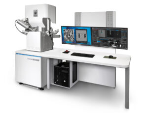
TESCAN AMBER 2
Versatile nanoanalytical FIB-SEM to expand your materials research capabilities.

TESCAN AMBER 2 X
A unique combination of Plasma FIB and fieldless UHR FE-SEM for multi-scale materials characterization.

TESCAN AMBER 2 CRYO
An alternative solution to prepare cryogenic slides for your TEM.

TESCAN CORETOM
O primeiro micro-CT dinâmico dedicado do mundo para as suas necessidades experimentais in situ.

TESCAN DYNATOM
O primeiro micro-CT dinâmico dedicado do mundo para as suas necessidades experimentais in situ.

TESCAN UNITOM
Multi-resolution micro-CT optimized for high throughput, diverse sample types and flexibility for your research