


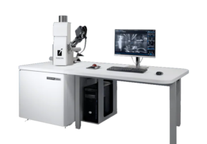

X-ray spectrometry is one of the most common techniques implemented in SEM for microanalysis. Energy-dispersive X-ray spectroscopy (EDX) and wavelength-dispersive X-ray spectroscopy (WDX) are two such techniques in which the characteristic of providing the elemental composition of the sample in the form of spectra (histograms) in which the individual elements that can be identified. The peaks in the EDX and WDX spectra correspond to X-ray lines characteristic of a specific element. Thus, spectra provide quantitative chemical characterization of samples.
In an EDX analysis, the entire energy range of characteristic X-rays is measured simultaneously, unlike WDX, where only a single wavelength (corresponding to one energy value) is measured at a time. Therefore, EDX analysis is faster compared to WDX.
In terms of power resolution, the WDX offers significantly better resolution than the EDX. Peaks missing in an EDX analysis are clearly resolved in a WDX spectrum. This is especially useful when analyzing trace elements.
For unknown samples, it is advisable to perform an initial EDX analysis that identifies the main elements present in the sample, followed by a more sensitive WDX analysis to resolve overlapping peaks and detect trace elements that exceed the detection limit of the EDX detector.
Electrons accelerated in the primary beam of a scanning electron microscope (SEM) can be diffracted by atomic layers in crystalline materials. These diffracted electrons can be detected and generate visible lines, called Kikuchi bands or “EBSPs” (electron backscatter patterns).
These patterns are effectively projections of the geometry of the lattice planes in the crystal and provide direct information about the crystal structure and crystallographic orientation of the grain from which they originate. Furthermore, together with the crystallographic parameters (orientation and their relationships) we will have the morphology (size and shape) and the chemical composition (phase identification).
To better meet your specific analytical needs, there are a variety of spectrometers that can be integrated into the TESCAN SEM and FIB-SEM systems.
Versatile UHR cryo-SEM for characterizing your biological and other beam-sensitive samples.
Analytical SEM for routine materials characterization, research, and micrometer-scale quality control applications.
Utilizamos cookies para melhorar a sua experiência no nosso site. Ao utilizar o nosso site, consente a utilização de cookies.
Gere abaixo as suas preferências de cookies:
Essential cookies enable basic functions and are necessary for the proper function of the website.
These cookies are needed for adding comments on this website.
Pode encontrar mais informações na nossa Cookie Policy e Política de privacidad.